Key Brain Structures Featured in Coloring Books
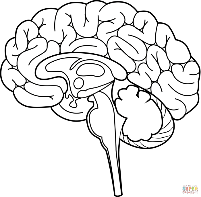
Brain anatomy coloring book answers – Brain anatomy coloring books offer a simplified yet effective way to learn about the complex structure of the human brain. By engaging visually with the different regions, learners can better understand their relative locations and appreciate the intricate organization of this vital organ. This section details the major brain regions commonly depicted in such books, along with their crucial functions.
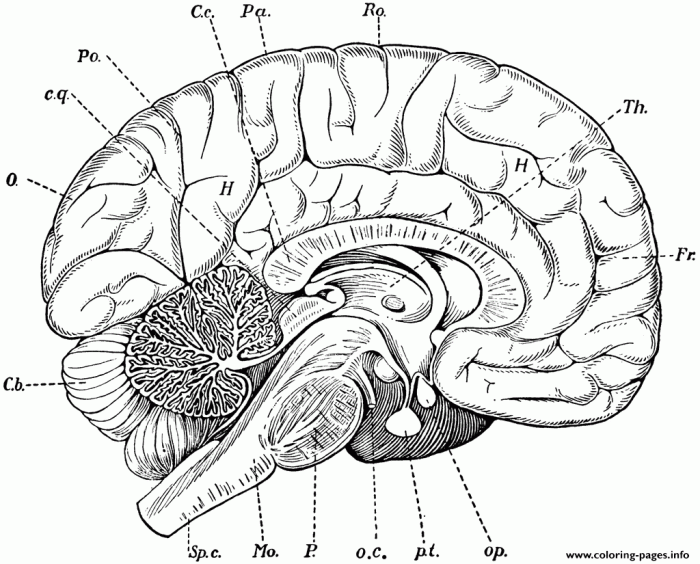
Coloring books typically focus on the most prominent and functionally distinct areas of the brain. While the level of detail varies, a core set of structures consistently appears, allowing for a foundational understanding of brain organization and function.
Major Brain Regions and Their Functions
The following table summarizes the key brain regions frequently found in brain anatomy coloring books, their general locations, and their primary functions. It is important to note that these functions are highly interconnected, and the brain operates as a holistic system, not as isolated modules.
| Brain Region | Location | Primary Functions | Illustrative Example |
|---|---|---|---|
| Cerebrum | Largest part of the brain; occupies the upper portion of the skull. | Higher-level cognitive functions, including thinking, learning, memory, language, and voluntary movement. Divided into four lobes (frontal, parietal, temporal, occipital) each with specialized roles. | Solving a complex math problem relies heavily on the cerebrum’s frontal lobe (planning and decision-making) and parietal lobe (spatial reasoning). |
| Cerebellum | Located at the back of the brain, beneath the cerebrum. | Coordination of movement, balance, and posture. Plays a role in motor learning and some cognitive functions. | Riding a bicycle requires the cerebellum to coordinate muscle movements for balance and smooth motion. |
| Brainstem | Connects the cerebrum and cerebellum to the spinal cord; located at the base of the brain. | Controls essential life functions such as breathing, heart rate, blood pressure, and sleep-wake cycles. | Automatic regulation of breathing while sleeping is a function of the brainstem. |
| Hypothalamus | Small region located below the thalamus, in the brainstem. | Regulation of body temperature, hunger, thirst, sleep, and the endocrine system (hormone production). | Feeling thirsty and seeking water is driven by the hypothalamus’s regulation of fluid balance. |
Analyzing Coloring Book Answers

Coloring books, while seemingly simple, offer a powerful tool for reinforcing learning, particularly in complex subjects like brain anatomy. By engaging students in active participation, they promote better retention and understanding compared to passive learning methods. Analyzing the completed coloring book pages allows educators to assess comprehension and identify areas needing further attention. This analysis will focus specifically on the accuracy of the coloring and labeling of the cerebrum’s lobes and their associated functions.
Cerebral Lobes and Their Functions
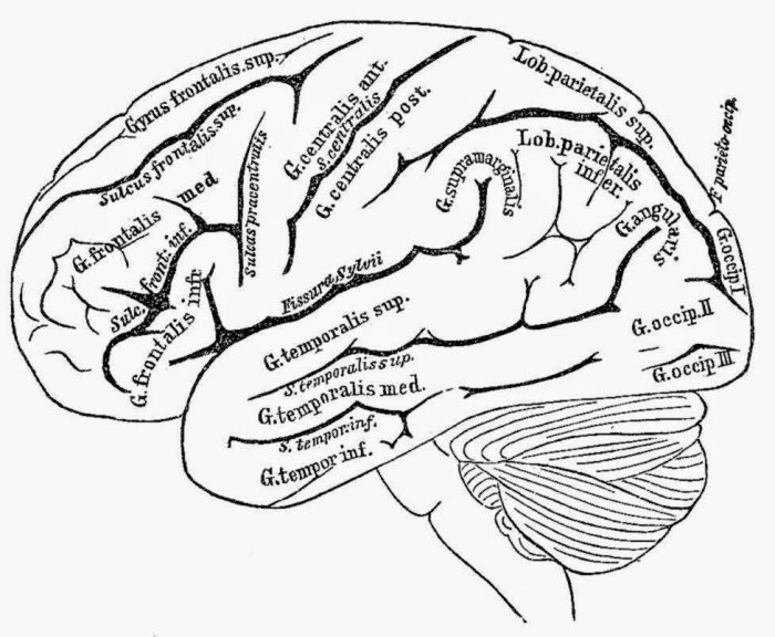
The cerebrum, the largest part of the brain, is divided into four lobes: frontal, parietal, temporal, and occipital. Each lobe plays a distinct role in cognitive functions, though they work interdependently. Understanding the unique contributions of each lobe is crucial for comprehending the brain’s overall functionality. Misconceptions regarding the specific functions of each lobe can lead to inaccurate interpretations of neurological conditions and limitations in effective treatment strategies.
Therefore, accurate coloring and labeling of these lobes are essential to demonstrate a solid understanding of basic brain anatomy.
Comparison of Cerebral Lobe Functions
The following table summarizes the key functions associated with each of the four lobes of the cerebrum. Note that these functions are not mutually exclusive, and many cognitive processes involve multiple lobes working together.
| Lobe | Primary Functions | Associated Cognitive Processes | Examples of Coloring Book Reinforcement |
|---|---|---|---|
| Frontal | Higher-level cognitive functions, voluntary movement, planning, decision-making, speech production (Broca’s area). | Problem-solving, working memory, attention, social behavior, personality. | Coloring a section representing the frontal lobe while simultaneously solving a simple puzzle or planning a route on a map provided alongside the coloring page. |
| Parietal | Processing sensory information (touch, temperature, pain, pressure), spatial awareness, navigation. | Spatial reasoning, object recognition, reading, writing. | Coloring a section representing the parietal lobe while simultaneously tracing a complex shape or identifying objects in a simple scene depicted on the page. |
| Temporal | Auditory processing, memory formation (hippocampus), language comprehension (Wernicke’s area). | Long-term memory, speech understanding, facial recognition. | Coloring a section representing the temporal lobe while simultaneously listening to a short auditory description of an object or event and then drawing it. |
| Occipital | Visual processing. | Object recognition, depth perception, color vision. | Coloring a section representing the occipital lobe while simultaneously identifying colors or shapes in a picture presented on the page. The act of coloring itself reinforces visual processing. |
Examples of Coloring Book Activities Reinforcing Cerebral Function Understanding
Coloring activities can effectively reinforce understanding of cerebral functions. For instance, a coloring page could depict a simplified brain with each lobe a different color, accompanied by labels and brief descriptions of their functions. Students could then color the lobes, reinforcing the visual association between the brain region and its role. More advanced activities could involve labeling specific areas within the lobes (like Broca’s and Wernicke’s areas) or matching functional descriptions to the corresponding lobe.
This interactive approach transforms passive learning into an active, engaging process, fostering better retention and a deeper understanding of brain anatomy. Furthermore, activities like associating a specific color with a particular function can aid in memorization and recall. For example, associating the color blue with language processing (temporal lobe) can help students remember the location and function of that area.
Illustrations and Visual Representations

Effective visual representation is crucial for a brain anatomy coloring book, ensuring comprehension and engagement. The illustrations must be both accurate and simplified to avoid overwhelming the user, particularly when dealing with complex structures and processes. The goal is to create visually appealing diagrams that clearly convey the key features and functions of each brain region.
Limbic System Visual Representation
The limbic system, responsible for emotion, memory, and motivation, can be illustrated as a ring of interconnected structures surrounding the brainstem. The amygdala, depicted as almond-shaped structures on either side of the thalamus, should be shown receiving input from various sensory areas and projecting to the hypothalamus and other limbic regions. Its function, processing emotional responses, particularly fear and aggression, can be subtly suggested through color coding or accompanying text.
The hippocampus, a seahorse-shaped structure nestled within the temporal lobe, should be illustrated with its characteristic curves and its close proximity to the amygdala. Its role in memory consolidation and spatial navigation can be highlighted through visual cues, perhaps using arrows to show its connections to other brain areas involved in memory processing. The interconnection between the amygdala and hippocampus should be clearly shown, indicating their collaborative role in emotional memory formation.
For instance, a visual connection could be represented by colored pathways linking the two structures.
Simplified Neuron and Synapse Representation
Illustrating the intricate network of neurons and synapses requires simplification for a coloring book. A single neuron can be depicted as a stylized cell with a cell body (soma), dendrites (branching input structures), and an axon (a long output fiber). Synapses, the junctions between neurons, can be represented as small, colored circles or dots at the end of the axon, where neurotransmitters are released.
Okay, so like, finding brain anatomy coloring book answers can be a total brain-buster, right? But if you’re, like, totally burnt out on that, check out this awesome big city greens coloring book for a chill break. It’s way more fun than memorizing the hippocampus, tbh. Then, once you’re all relaxed, you can totally crush those brain anatomy answers!
To visually represent the network, multiple neurons could be shown interconnected, with colored synapses highlighting the pathways of information flow. The coloring activity itself could involve coloring different types of neurons or different neurotransmitters to enhance learning and engagement. This simplified representation, while omitting many intricate details, successfully conveys the basic principles of neuronal communication.
Spinal Cord Cross-Section
A cross-section of the spinal cord should clearly distinguish between the grey matter and the white matter. The grey matter, shaped like a butterfly or “H,” should be centrally located and colored differently from the surrounding white matter. The dorsal horns (receiving sensory information) and the ventral horns (sending motor commands) should be clearly labeled and differentiated within the grey matter.
The white matter, surrounding the grey matter, should be shown as tracts of myelinated axons, potentially using different shades to represent ascending (sensory) and descending (motor) pathways. This visual representation should emphasize the role of the grey matter in processing information and the role of the white matter in transmitting information up and down the spinal cord. Simple labels indicating sensory and motor functions associated with the dorsal and ventral horns, respectively, will further enhance understanding.
For example, a simple legend could correlate specific colors with sensory or motor functions.
Advanced Concepts and Applications: Brain Anatomy Coloring Book Answers

Creating accurate and engaging brain anatomy coloring books presents significant challenges. The complexity of the brain, with its intricate network of interconnected structures and subtle variations, necessitates careful simplification for a target audience likely unfamiliar with advanced neuroanatomy. Successfully navigating this simplification while maintaining educational value requires thoughtful design and pedagogical considerations.The process of translating the three-dimensional structure of the brain into a two-dimensional coloring book format inherently involves compromises.
Fine details are lost, and the spatial relationships between structures may be distorted. The choice of which structures to include and the level of detail provided requires careful consideration of the learning objectives and the age and background of the intended users. For example, a coloring book aimed at young children might focus on the major lobes of the brain, while a book for older students could include more detailed representations of subcortical structures like the basal ganglia or the hippocampus.
Color-Coding for Differentiation of Brain Regions and Functions
Effective color-coding is crucial for differentiating brain regions and their associated functions in a coloring book. A consistent color scheme should be established, ideally using colors associated with specific functions or regions in established neuroanatomical literature. For example, the frontal lobe could consistently be represented in blue to associate it with executive functions, while the occipital lobe could be represented in red to represent its role in visual processing.
A key or legend should accompany the coloring pages to explain the color-coding scheme and provide a concise description of each region’s function. Using a color wheel or similar visual aid to demonstrate the relationships between brain regions and functions could further enhance understanding. Furthermore, varying the shades of a color can help differentiate sub-regions within a larger brain area.
For instance, different shades of blue could be used to delineate Broca’s area and Wernicke’s area within the frontal and temporal lobes, respectively, highlighting their distinct roles in language processing.
Engaging Learning Through Coloring Book Activities, Brain anatomy coloring book answers
Coloring book activities can be made significantly more engaging and memorable by incorporating interactive elements. Simple quizzes or labeling exercises integrated into the coloring pages can reinforce learning. For instance, a coloring page of the cerebellum could include blank labels for key structures like the vermis and cerebellar hemispheres, requiring the user to identify and label these components after coloring the page.
Adding puzzles, mazes, or other games related to brain function and anatomy can enhance the learning experience and cater to diverse learning styles. Furthermore, the inclusion of interesting facts or anecdotes about each brain region can spark curiosity and make the learning process more enjoyable. For example, a coloring page depicting the amygdala could include a brief description of its role in processing emotions, and a related fact about the amygdala’s involvement in fear responses.
This approach fosters a more holistic and engaging understanding of the brain’s intricate workings, transforming a simple coloring activity into a more effective learning tool.
Quick FAQs
What age group are brain anatomy coloring books suitable for?
Brain anatomy coloring books can be adapted for various age groups, from elementary school children (with simplified illustrations and basic information) to high school and college students (with more complex details and advanced concepts).
Are there different levels of difficulty in brain anatomy coloring books?
Yes, many coloring books offer varying levels of complexity, catering to different levels of understanding and knowledge. Some focus on basic structures, while others delve into more intricate details.
Where can I find brain anatomy coloring books?
Brain anatomy coloring books can be found online through various retailers, educational suppliers, and online bookstores. Many are also available in libraries and educational institutions.
How can I use a brain anatomy coloring book most effectively?
For optimal learning, use the coloring book in conjunction with other learning materials, such as textbooks or online resources. Take your time, focus on understanding the structures and functions, and actively engage with the information presented.
